WhatsApp!
+91 704 548 7309

Uterine Fibroid
A uterine fibroid is a leiomyoma (benign tumor from smooth muscle tissue) that originates from the smooth muscle layer (myometrium) of the uterus.
The malignant version of a fibroid is extremely uncommon and termed a leiomyosarcoma.
Fibroids are the most common benign tumors in females and typically found during the middle and later reproductive years. While most fibroids are asymptomatic, they can grow and cause heavy and painful menstruation, painful sexual intercourse, and urinary frequency and urgency. Some fibroids may interfere with pregnancy although this appears to be very rare.
Signs and symptoms
Fibroids, particularly when small, may be entirely asymptomatic. Symptoms depend on the location of the lesion and its size. Important symptoms include abnormal gynecologic hemorrhage, heavy or painful periods, abdominal discomfort or bloating, painful defecation, back ache, urinary frequency or retention, and in some cases, infertility. During pregnancy they may also be the cause ofmiscarriage, bleeding, premature labor, or interference with the position of the fetus.
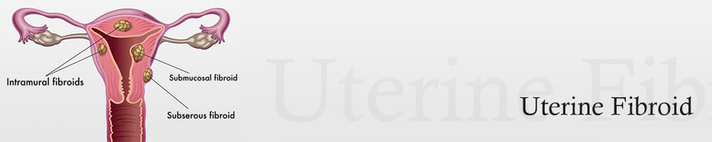
Location and classification
Schematic drawing of various types of uterine fibroids: a=subserosal fibroids, b=intramural fibroids, c=submucosal fibroid, d=pedunculated submucosal fibroid, e=fibroid in statu nascendi, f=fibroid of the broad ligament.
Growth and location are the main factors that determine if a fibroid leads to symptoms and problems.[3] A small lesion can be symptomatic if located within the uterine cavity while a large lesion on the outside of the uterus may go unnoticed.
Fibroids may be single or multiple. Most fibroids start in an intramural location, that is the layer of the muscle of the uterus. With further growth, some lesions may develop towards the outside of the uterus or towards the internal cavity. Secondary changes that may develop within fibroids are hemorrhage, necrosis, calcification, and cystic changes.

Pathogenesis
Known risk factors are African descent, nulliparity, obesity, polycystic ovary syndrome, diabetes and hypertension.
Diagnosis
While a bimanual examination typically can identify the presence of larger fibroids, gynecologic ultrasonography (ultrasound) has evolved as the standard tool to evaluate the uterus for fibroids. Sonography will depict the fibroids as focal masses with a heterogeneous texture, which usually cause shadowing of the ultrasound beam. The location can be determined and dimensions of the lesion measured. Also magnetic resonance imaging (MRI) can be used to define the depiction of the size and location of the fibroids within the uterus.

A very large (9 cm) fibroid of the uterus which is causingpelvic congestion syndrome as seen on CT |

A very large (9 cm) fibroid of the uterus which is causing pelvic congestion syndrome as seen on ultrasound |

A relatively large submucosal leiomyoma; it fills out the major part of the endometrial cavity |

A small uterine fibroid seen within the wall of themyometrium on a cross sectional ultrasound view |

Two calcified fibroids (in the uterus) |

thumb|A subserosal uterine fibroid with a diameter of 5 centimeters |
Coexisting disorders
Fibroids that lead to heavy vaginal bleeding lead to anemia and iron deficiency. Due to pressure effects gastrointestinal problems such as constipation and bloatedness are possible. Compression of the ureter may lead to hydronephrosis. Fibroids may also present alongside endometriosis, which itself may cause infertility.
In very rare cases, malignant (cancerous) growths, leiomyosarcoma, of the myometrium can develop.[38] In extremely rare cases uterine fibroids may present as part or early symptom of the hereditary leiomyomatosis and renal cell cancersyndrome.
Treatment
Most fibroids do not require treatment unless they are causing symptoms. After menopause fibroids shrink and it is unusual for fibroids to cause problems.
Symptomatic uterine fibroids can be treated by :
- medication to control symptoms
- medication aimed at shrinking tumours
- ultrasound fibroid destruction
- myomectomy or radio frequency ablation
- hysterectomy
- uterine artery embolization
Myomectomy

submucosal fibroid in hysteroscopy |

Treatment of an intramural fibroid bylaparoscopic surgery |

After treatment of an intramural fibroid bylaparoscopic surgery |
Myomectomy is a surgery to remove one or more fibroids. It is usually recommended when more conservative treatment options fail for women who want fertility preserving surgery or who want to retain the uterus.
There are three types of myomectomy :
- hysteroscopic myomectomy
- laparoscopic myomectomy
- laparotomic myomectomy
Hysterectomy
Hysterectomy was the classical method of treating fibroids. Although it is now recommended only as last option.